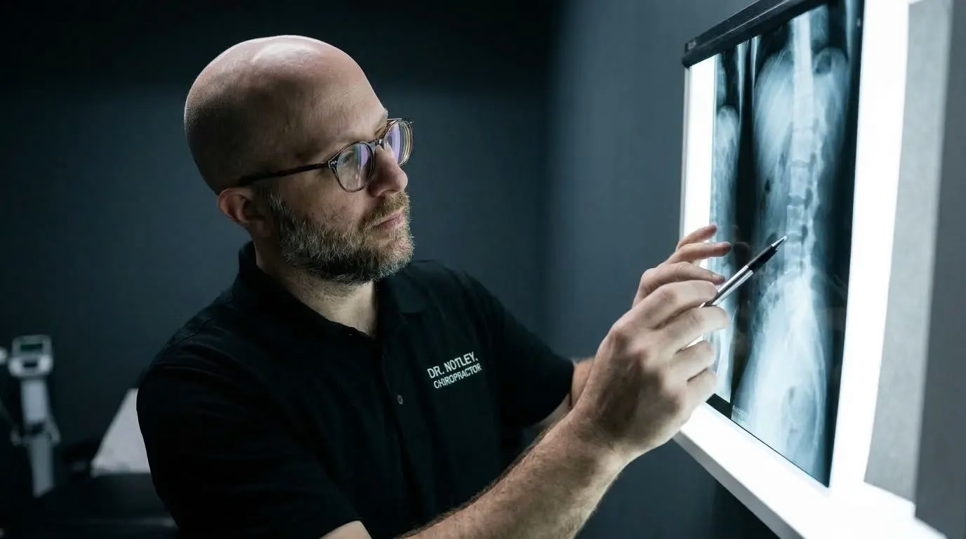
What Is Disc Desiccation? (And Why It’s Just Grey Hair for Your Spine)
The fear: “My spinal discs are drying up, rotting, and dying.”
Here is the more accurate version. Your discs are doing exactly what skin, cartilage, and connective tissue all over your body do over the decades. They gradually hold a little less water. It is not decay. It is chemistry.
Each disc has a gel-like center (the nucleus pulposus) held in place by tough, ring-like fibers (the annulus fibrosus). That gel center is rich in proteoglycans, molecules that act like tiny sponges, pulling water in and letting the disc handle load and stay cushioned. As we age, the number and quality of those proteoglycans gradually decline, so the disc holds less water than it did at 20. On an MRI, that shows up as a “dark disc” on certain sequences, which is exactly what radiologists are describing when they write “desiccation.”
pain-free 20-year-olds already show some disc degeneration on MRI — climbing toward near-universal by the 70s and 80s. It’s a weak, mostly silent finding, not a diagnosis.
Here is the detail that gets left out of most radiology reports, and it is the useful one. Intervertebral discs have no direct blood supply. The cells inside get most of their nutrients, things like oxygen and glucose, by diffusion. These small molecules seep in from tiny blood vessels at the edge of the disc, and waste products seep back out the same way.
On top of that slow, steady diffusion, there is a second process that depends on movement. Every time you load and unload your spine, by walking, shifting position, standing up, or lying down, fluid moves in and out of the disc. Stand up and a little fluid squeezes out. Lie down and it draws back in. This day-to-day fluid exchange, sometimes called imbibition, helps keep the whole system flushed and hydrated.
That is why the discs that stay healthiest over a lifetime are generally not the ones protected from all movement. They are the ones that keep moving regularly. Long periods of static sitting, or guarding your back “just in case,” actually reduce this natural exchange. Studies show that sustained, still loading slows the movement of nutrients into the disc, and that long-term immobilization can drive degeneration rather than prevent it. Ironically, the instinct to freeze up and protect a “drying” disc can work against the very processes that keep it resilient. (For more on how load-bearing activity affects disc health specifically, see Can Running Actually “Heal” Your Spine?)
Move often, not hard
Frequent position changes throughout the day matter more than one long workout.
Walking is disc food
Gentle, repetitive loading keeps fluid and nutrients moving — see our WalkBack Program for a structured way to build this in.
Don’t fear bending
Controlled bending and extending, introduced gradually, is part of how discs stay adaptable.
Build capacity, not fragility
A graded strength and mobility program does more for a “desiccated” disc than rest ever will.
Disc desiccation is a normal marker of a spine that has been alive and active for a while, not a warning label. If desiccation is one of several findings on your report, or your pain has stuck around, our full guide to discogenic lower back pain covers recovery timelines and treatment options in more depth. If you want a movement plan built around what your spine can actually handle right now, that is exactly what an assessment is for.
Why Words Matter: The Science of the “Threat Dial”
How your MRI is explained to you can actually change how fast you recover.
In a 2021 randomized trial in the European Spine Journal, Rajasekaran and colleagues split back pain patients into two groups getting identical physical treatment. One group received a raw, line-by-line breakdown of every scary-sounding finding. The other had the same findings explained as normal, age-related change.
The group given the plain, reassuring explanation came away far less alarmed about their spine and less convinced they needed surgery, while the group handed the standard report were left more distressed by the very same images.
“Same scans, same treatment, different words.”
This is not a one-off. Other research has found that the specific labels used—words like “degeneration,” “arthritis,” and “bulge”—lower people’s expectations of recovery and raise their belief that surgery is needed, compared with plainer terms. Patients who absorb phrases like “wear and tear” or “crumbling” tend to report a worse outlook on their own recovery, independent of what the scan actually shows.
Pain science gives us a clean way to understand why. Pain is an output, not a readout. Your nervous system does not measure damage directly. It makes a best guess about threat, built from tissue signals plus your beliefs, your mood, and everything you have been told about your body. That is why the same disc finding can be silent in one person and severely painful in another. The structure is not different. The threat dial is.
Moving Beyond the Image: Calm the Alarm, Build the Buffer
An MRI shows what your spine looks like lying perfectly still inside a magnet. It does not show how well your joints move, how strong your core is, or how resilient your body can become. And as the research above shows, it is a weak enough signal on its own that it should not set your expectations by itself.
A good, evidence-based plan works in two phases:
Calm the Alarm
When acute pain strikes, targeted joint mobilization, manual therapy, and myofascial release help settle the nervous system, ease protective muscle guarding, and restore pain-free joint movement.
Build the Buffer
Progressive rehabilitation—core control, hip mobility, gradual strength loading—expands your physical capacity. This is where athletic therapy bridges the gap, keeping daily life well below your sensitivity threshold.
Your MRI report is a tool to rule out rare emergencies and add useful context. It is not a life sentence, and it does not dictate your future. Your spine is far more adaptable than the report makes it sound.
Book a Movement Assessment in Winnipeg
If desiccation is one of several findings on your report, or your pain has stuck around, our full guide to discogenic lower back pain covers recovery timelines and treatment options in more depth.
If you want a movement plan built around what your spine can handle right now, that is exactly what an assessment is for. Dr Notley sees patients at two Winnipeg locations — St. Vital and Downtown. No referral needed.
Book an AppointmentSources and Further Reading
The claims in this article are drawn from peer-reviewed research.
View all 14 references
- Brinjikji W, et al. Systematic literature review of imaging features of spinal degeneration in asymptomatic populations. AJNR Am J Neuroradiol. 2015.
- Brinjikji W, et al. MRI findings of disc degeneration are more prevalent in adults with low back pain than in asymptomatic controls: a systematic review and meta-analysis. AJNR Am J Neuroradiol. 2015.
- Kasch R, et al. Association of lumbar MRI findings with current and future back pain in a population-based cohort study. Spine. 2022.
- Dragsbæk L, et al. Associations between sum scores or combinations of MRI findings in the lumbar spine and low back pain-related outcomes: a systematic review. Eur J Pain. 2025.
- Dragsbæk L, et al. Associations between single and combined lumbar MRI findings and low back pain with and without leg pain: a cross-sectional study. BMC Musculoskelet Disord. 2026.
- Rajasekaran S, et al. The catastrophization effects of an MRI report on the patient and surgeon and the benefits of ‘clinical reporting’: results from an RCT and blinded trials. Eur Spine J. 2021.
- O’Keeffe M, et al. Public and patient perceptions of diagnostic labels for non-specific low back pain: a content analysis. Eur Spine J. 2022.
- Rashed S, et al. Systematic review and meta-analysis of predictive factors for spontaneous regression in lumbar disc herniation. J Neurosurg Spine. 2023.
- Urban JPG, Smith S, Fairbank JCT. Nutrition of the Intervertebral Disc. Spine (Phila Pa 1976). 2004.
- Belavý DL, et al. Can Exercise Positively Influence the Intervertebral Disc? Sports Med. 2016.
- Gullbrand SE, et al. Dynamic Loading-Induced Convective Transport Enhances Intervertebral Disc Nutrition. Spine (Phila Pa 1976). 2015.
- Arun R, et al. What Influence Does Sustained Mechanical Load Have on Diffusion in the Human Intervertebral Disc? (2009 ISSLS Prize Winner). Spine (Phila Pa 1976). 2009.
- Che YJ, et al. Intervertebral disc degeneration induced by long-segment in-situ immobilization. BMC Musculoskelet Disord. 2018.
- Samanna CL, et al. The Impact of Exercise on Intervertebral Disc Health: A Systematic Review and Meta-Analysis. Sports Med. 2026.
What does “disc desiccation” mean on an MRI?
Disc desiccation is the natural loss of moisture in your spinal discs over time. It is a normal biological change, much like grey hair or skin wrinkles, and it is common in people with zero back pain.
Can a disc bulge or herniation heal on its own?
Often, yes, and the more dramatic-looking herniations are actually the ones most likely to shrink. As the body’s immune system clears away displaced disc material, larger extruded and “sequestered” herniations frequently resorb, while a plain bulge changes less on imaging. Either way, symptoms usually improve over months with conservative care, and the size of a finding does not decide your outcome.
Does facet joint hypertrophy mean I have severe arthritis?
No. It means the small stabilizing joints in your spine have adapted and thickened to handle load. It can contribute a little to pain that flares under stress, but it is a sign of adaptation, not active decay, and it is not a guarantee of chronic pain.
I have several findings on my MRI. Does that mean I’m in worse shape?
It is worth taking a bit more seriously than a single finding, but it is still not a verdict. Research shows a gradual link between the number of findings and the amount of pain and disability people report. Real, but modest, and heavily shaped by how strong and mobile the muscles and joints around your spine are. Several findings are a reason to build capacity deliberately, not a reason to assume the worst.
Do I need an MRI before starting rehab or hands-on care?
In most cases, no. Unless you have clinical “red flags,” such as progressive leg weakness, numbness in the saddle or groin area, changes in bowel or bladder control, significant trauma, unexplained weight loss, or fever, a physical movement assessment is far more useful for building an effective, personalized recovery plan. If any of those red flags are present, see a clinician promptly.