
Modic Changes MRI: What Your Spinal Bone Marrow is Doing
Have you ever looked at an MRI report and seen unfamiliar terms that instantly made your heart sink? You’re certainly not alone. Radiology reports are often filled with complex medical jargon, and one phrase that frequently causes worry is “Modic changes.”
The fear
Modic changes aren’t your bones rotting.
“My spinal bones are deteriorating or rotting.”
“Modic changes.” Just seeing those words on your MRI report can feel confusing, even alarming. It sounds highly medical, like your very bones are actively decaying or facing some mysterious disease. It’s completely natural for that phrase to spark fear and make you worry about the integrity of your spine.
But here’s the reassuring truth: much like other intimidating terms we’ve explored (like Disc Desiccation or Bone Spurs), “Modic changes” describes something far less dramatic.
Side view of two stacked vertebrae with a disc between them. The orange band marks where Modic changes appear. Simplified illustration.
They’re named after Dr. Michael Modic, who first described them in 1988.
Your bones are always remodeling
Normal adaptation, not decay
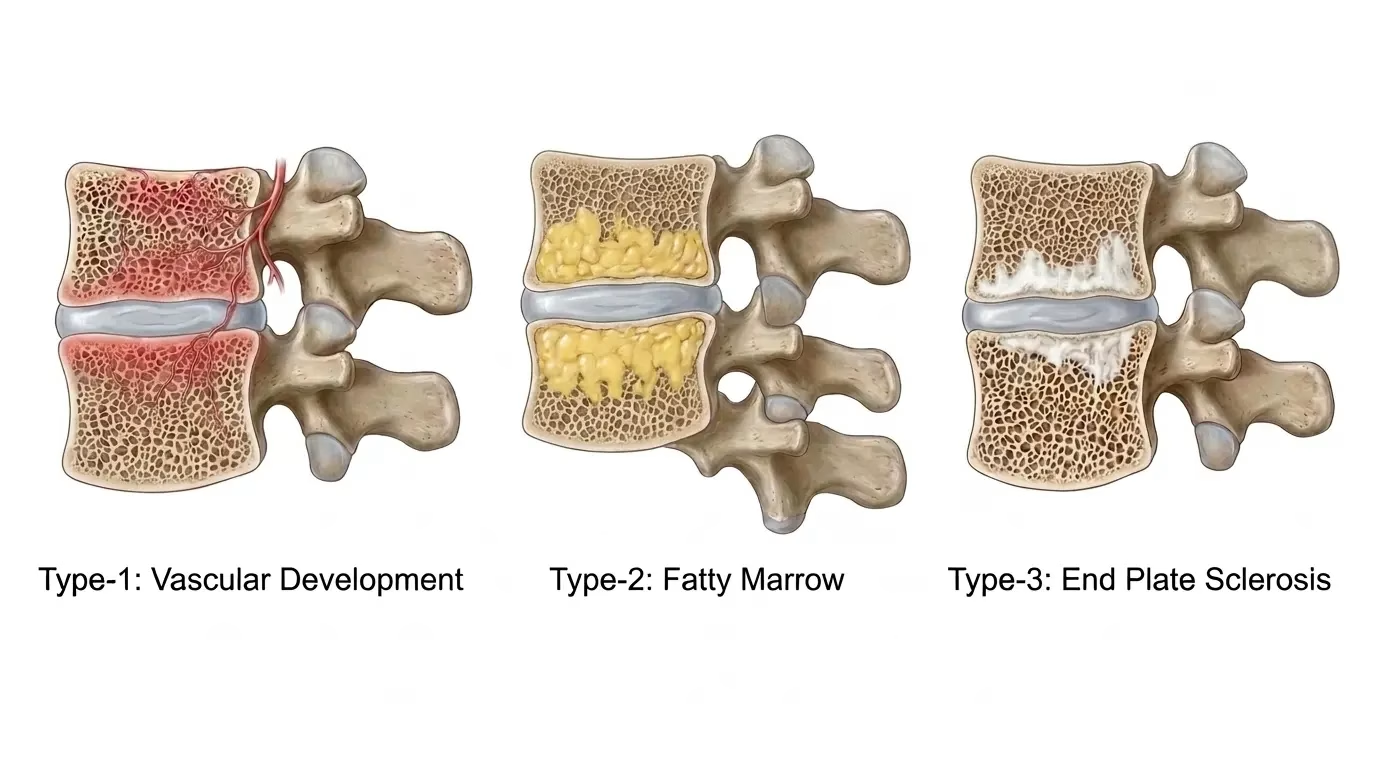
Think of your bones not as static structures, but as living, dynamic tissue. They’re constantly rebuilding, adapting, and responding to the loads and forces they experience every day. Modic changes show us how your bone is reacting to changes in disc health and how weight is being distributed across your spine.
There are three main types. Each looks different on the two main kinds of MRI image, called T1 and T2.
The “Hot Spot”
Bright on T2-weighted scans. It indicates a phase of active inflammation and shifts in blood supply within the bone marrow.
If you have new back pain, Type 1 is the one most often associated with that temporary sensitivity, like a “hot spot” as the bone remodels. It doesn’t mean damage, but it might mean irritation.
The “Settling Down” Phase
Bright on T1-weighted scans. It reflects fatty changes in the bone marrow and often follows a Type 1 phase.
We generally see this as a more stable, long-term adaptation, less commonly linked to active pain than Type 1. Your body has moved past the initial irritation.
The “Stronger” Phase
Dark on both T1 and T2 scans. It indicates bone sclerosis, meaning the bone has hardened and thickened.
This is a sign of long-term adaptation and repair, usually a stable, less painful finding. Your body has reinforced the area.
Schematic of how each type tends to look on a scan: dark squares are dark signal, light squares are bright signal. Not real MRI images.
These different changes are all part of your body’s attempt to adapt and stabilize your spine. Research shows they are common, and Type 1 in particular can change over time, sometimes converting to Type 2.
What Modic changes mean for your pain
One piece of the puzzle
Sometimes associated with temporary back pain and sensitivity, which makes sense given the active inflammatory process in the bone. If you have Type 1 changes and new back pain, it’s a useful piece of information that can help guide your treatment plan.
Modic changes are found in people with no back pain. Like a disc bulge or facet joint osteoarthritis, their presence on a scan doesn’t automatically mean they’re the cause of your specific symptoms.
It’s one piece of the puzzle that needs to be considered alongside your clinical examination: how you move, and what your body tells us.
What actually helps
Support the body’s natural healing
Even if Type 1 changes are contributing to your pain, it doesn’t mean your bones are “rotting” or that you need aggressive interventions. The focus is on supporting natural healing, calming active inflammation, and promoting healthy spinal mechanics.
Avoid excessive immobilization
A little rest can feel good during acute pain, but prolonged inactivity can slow healthy bone remodeling. Gentle, progressive movement helps nourish your discs and bones.
Targeted hands-on care (chiropractic)
Chiropractic care helps reduce nervous system threat and ease protective muscle guarding, creating a safe window for comfortable movement and less local irritation.
Progressive load and movement (athletic therapy)
Gradually strengthening your core and improving hip mobility helps distribute load across your spine, encouraging healthy bone adaptation and expanding your resilience.
Anti-inflammatory strategies
Not a magic cure, but good nutrition, adequate sleep, and stress management support your body’s natural anti-inflammatory processes, which can help calm Type 1 changes.
Your bones are resilient and constantly capable of remodeling and adapting. Understanding Modic changes means seeing them as an adaptive process, your body trying to heal and reinforce, not a sentence of decay. With the right approach, you can manage any associated temporary pain and build a stronger, more confident spine.
Want help making sense of your own report?
Book an assessment with Winnipeg Chiropractor & Athletic Therapist Dr. Chris NotleyPart of our MRI Translation Guide series
More from the series
Sources and further reading (4)
- Modic MT, et al. Imaging of degenerative disk disease. Radiology. 1988. (Original description of Modic changes.)
- Kjaer P, et al. Modic changes and their associations with clinical symptoms: a systematic review. BMC Musculoskelet Disord. 2018. (Systematic review on association with pain.)
- Herlin T, et al. Modic changes in the lumbar spine: prevalence and association with low back pain. Eur Spine J. 2018. (Prevalence and associations.)
- Jensen TS, et al. Modic changes: a systematic review. Spine. 2012. (Comprehensive review.)