
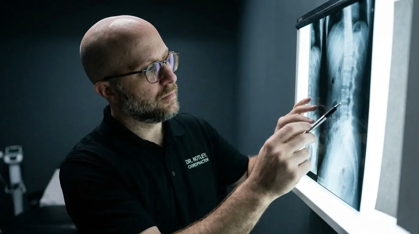
Explaining MRI/X-ray to Patients: Your Guide to Radiology Reports
If you have ever read your MRI or X-ray report after a bad bout of back pain, you know the feeling that comes next. Words like “disc desiccation,” “loss of disc height,” “annular tear,” or “facet joint hypertrophy” jump off the page in cold, clinical black and white.
It is completely natural to read those terms and panic. You picture your spine crumbling, your back permanently damaged, one wrong twist away from a wheelchair.
Here is what most people are never told: radiology reports are not written for you. They are written by radiologists for surgeons and specialists, in precise anatomical language built to rule out rare emergencies. They describe the structure of your spine. On their own, individual findings are weak, easy to overweight clues. They are not a verdict on your pain, your strength, or your ability to heal.
Understanding what your lumbar MRI actually says, and where it fits in the bigger picture, is often the first real step toward a confident recovery.
Two Stories, and Neither One Is the Whole Truth
For years, spine care ran on a simple idea. Find something on the scan, name it, treat it. A bulge or a bit of lost disc height became “the” diagnosis.
Then research pushed back hard, and rightly so. Those same “abnormal” findings turned out to show up constantly in people who have never had a day of back pain in their life. The message flipped to: “MRI findings mean nothing. Everyone has degeneration.”
That correction did real good. It talked a lot of frightened people off the ledge. But it overshot. The honest picture sits between the two slogans. Structural changes are common and rarely dangerous on their own, and they are still real, if modest, pieces of information, especially when several of them show up together. This guide holds both of those at once.
The “Wrinkles on the Inside” Rule
To make sense of your MRI, it helps to know how common these changes are in people who feel completely fine.
In a landmark 2015 review in the American Journal of Neuroradiology, Brinjikji and colleagues looked at lumbar MRIs from more than 3,000 people with zero back pain. The numbers reshaped how we read spinal imaging:
Disc degeneration without any back pain
Imaging studies of pain-free people show these changes show up on their own, often well before any pain does — and they get more common with age.
Source: Brinjikji et al., American Journal of Neuroradiology, 2015 — imaging findings in people with no back pain. These changes are common signs of aging, not proof that a disc is the cause of pain.
So finding a bulge or some degeneration on your scan is not, by itself, a red flag. Think of it as a “wrinkle on the inside” — a normal adaptation to decades of standing, walking, running, and carrying load, the same way grey hair and skin wrinkles show up without anything being wrong.
That said, a companion 2015 analysis from the same group compared people with back pain to people without, and found these same findings genuinely were more common in the painful group. Bulges, extrusions, and certain disc changes turned up noticeably more often. So a finding is not meaningless. It is just a weak signal on its own — real, but easy to overweight when you are staring at it alone on a page.
One Wrinkle vs. a Weathered Map
Here is the part that usually gets left out, and it is the part that actually helps you read your own report.
A single finding — one bulge, one level of mild degeneration — barely moves the needle. Research following thousands of people has found that even a fairly long list of individual findings adds less than a single point on a 0-to-10 pain scale. Real, but small.
The picture gets more meaningful when findings start to stack up. Newer research pooling dozens of studies has found a consistent pattern: the more findings present, and the more severe they are, the stronger the link to back pain and disability. Not an on-off switch, but a gradual dose-response curve. People with four or more distinct findings tend to report more pain than people with only one or two, though the difference is often modest and gets less predictable the more you zoom in.
So the honest way to read a busy report is not “my spine is wrecked,” and not “none of this matters.” It is closer to this: a spine with several findings may simply be working with a narrower margin — a quieter, more loaded system, not a broken one. That is useful information for building a recovery plan. It is not a diagnosis of doom.
Decoding your radiology report
Radiology language is written for other clinicians, not for the person reading it at home. Here is what five common phrases actually mean.
“My discs are drying up, rotting, and dying.”
A natural hydration shift in the disc over time, much like how skin loses some moisture and elasticity with age. On its own, it’s a normal age-related change, not structural decay.
“My disc popped out of place and is crushing my spinal cord.”
A slight outward spread of the disc’s flexible outer wall. The disc stays anchored to the vertebrae above and below, so it doesn’t “slip” out of place. Bulges are common in millions of active, largely pain-free adults.
“My spine is bone-on-bone and rotting away with arthritis.”
The small stabilizing joints behind your spine have thickened slightly to handle daily load, closer to a callus forming on your hands than structural failure. It can play a small part in pain that flares under stress, but on its own it’s adaptation, not collapse.
“My spinal shock absorber is completely gone.”
A minor narrowing between two vertebrae. Your body adapts by shifting load to the surrounding muscles and ligaments, which can be trained to be strong and resilient.
“My spinal nerves are being pinched in a vice.”
The small exit channels where nerves leave the spinal column have narrowed somewhat. Nerve irritation from a temporary flare-up often matters more than the physical space visible on a still image.
Ready to build a resilient, pain-free spine?
If you are in the Winnipeg area and want an honest, evidence-based assessment of your back pain, one that looks at the whole picture rather than a scan on a screen, schedule a consultation today.